
Analysis Of?Class I Malocclusion with Bimaxillary Protrusion.
- Age:26
- Gender:Female
- Chief complaint:Mouth protrusion, misaligned dental arches.
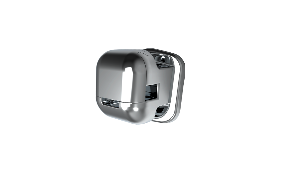
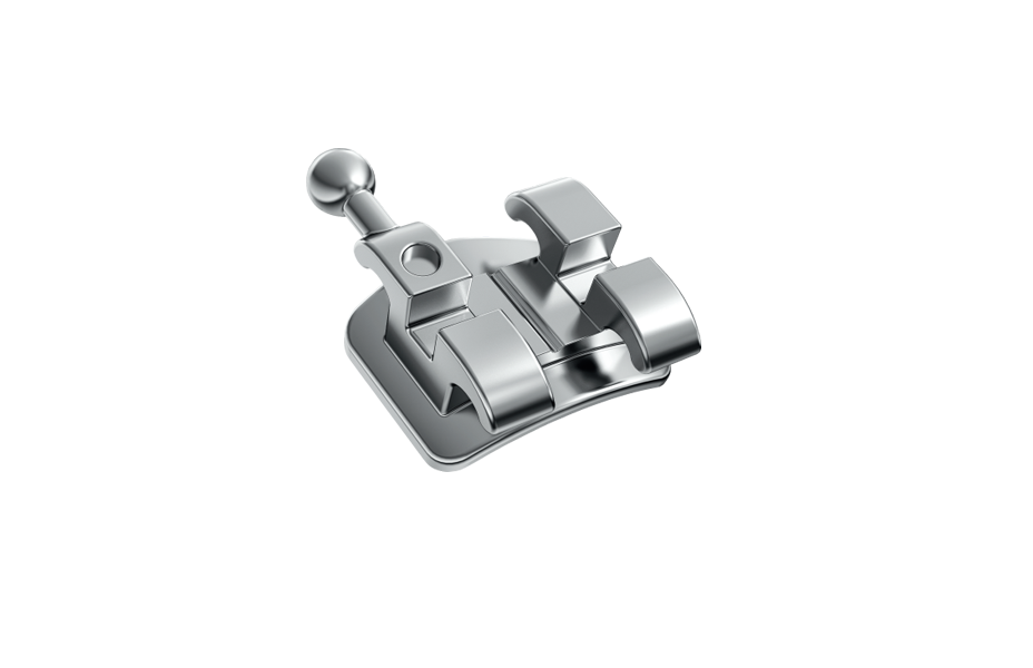
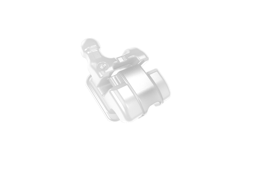
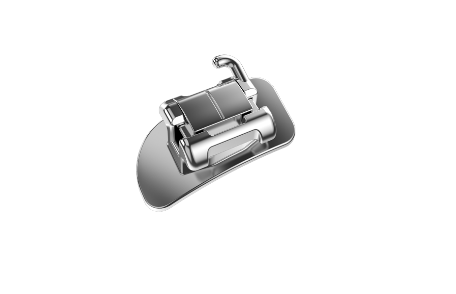
- Use product:PT V self-ligating brackets
- Treatment cycle:19 months
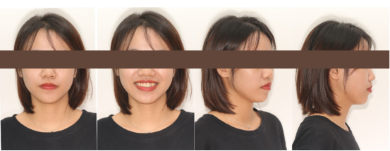
Facial Examination
Frontal view: Fuller on the left side than the right.
Lower one-third facial height ratio: Equal.
Tight lip closure.
Lateral view: Sagittal facial profile: Convex profile, protrusion of the upper lip, and outward rotation of the lower lip.
Nasolabial angle acute, tight lip closure.
Retruded lower jaw. Vertical facial profile: Balanced angles.
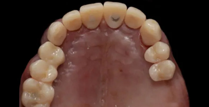
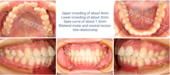
Intraoral Examination
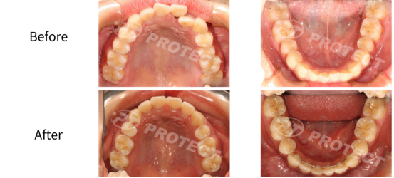
Upper crowding of about 4mm
Lower crowding of about 3mm
Spee curve of about 1.5mm
Bilateral molar and neutral incisor bite relationship
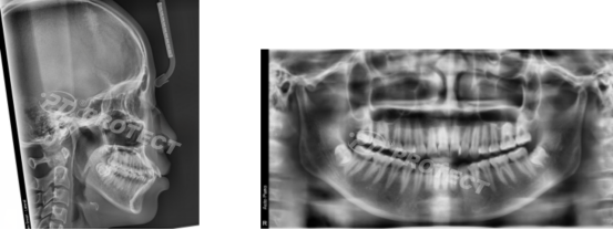
Pre-Orthodontic Head Measurement and Analysis
Bilateral condyles are relatively symmetrical.
Upper jaw sinus positions are essentially normal.
Bilateral mandibular ramus heights are asymmetrical, with the right side longer than the left side.
Teeth 11 and 21 have received root canal treatment.
Diagnosis:
1. Convex facial profile with protruding upper and lower lips, and an acute nasolabial angle.
2. Class I skeletal relationship with slight mandibular retrusion.
3. Anterior teeth exhibit lip protrusion.
Key Issues
1. Appearance (Front View): The lower one-third of the face is asymmetrical, with tension in lip closure. Gingiva is exposed during a big smile.
2. Appearance (Profile View): Class I skeletal relationship with an increased lower anterior facial height, convex facial profile, and tight chin muscles. Retruded mandible.
3. Molar and canine relationships are essentially in neutral occlusion, with upper and lower anterior teeth leaning.
Treatment Goals
1. To perform extraction treatment to correct the disguised presentation. Utilize the extraction spaces and implant-supported screws to improve the vertical relationship between the upper and lower jaws, ameliorating the convex profile.
2. Establish a neutral molar and canine occlusion to position the anterior teeth according to the smile arc and smile line.
3. Through orthodontic control of the occlusal plane, reduce the mandibular plane angle, achieve a counterclockwise rotation of the mandible, decrease excessive chin muscle tension, and enhance the soft tissue profile of the chin area.
Treatment Plan
1. Extract teeth 14, 24, 34, and 44. Utilize fixed 3rd generation PROTECT appliances for maxillary retraction to improve the convex profile.
2. Position the upper anterior teeth to restore the patient's smile arc as a reference, enhancing the smile arc and smile line.
3. Achieve the goal of 2mm vertical retraction of the upper anterior teeth with respect to the lower lip's margin.
4. Close the extraction spaces in the upper and lower arches to establish a neutral molar and canine occlusion.
5. Prescribe upper and lower clear retainers with a Hawley retainer. Full-time wear is required for the first 12 months, followed by nighttime wear.
Treatment Process
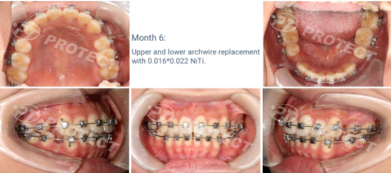
Month 6-Upper and lower archwire replacement with 0.016*0.022NiTi.
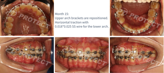
Month 15- Upper arch brackets are repositioned. Horizontal traction with 0.018*0.025 SS wire for the lower arch.
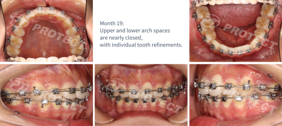
Month 19- Upper and lower arch spaces are nearly closed, with individual tooth refinements.
Intraoral Photos At The End Of The Treatment
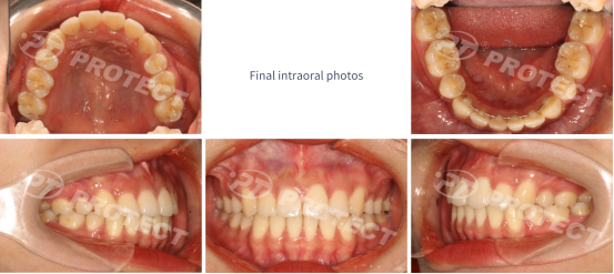
Final Intraoral Photos
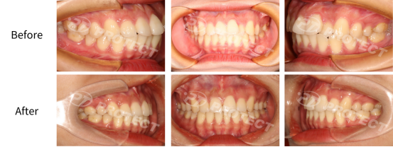
1. Anterior teeth exhibit a Class I relationship with neutral overbite and overjet.
2. Good matching of maxillary and mandibular dental arch forms.
3. Alignment of the midlines.
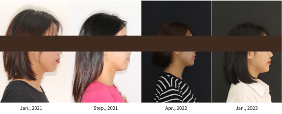
Postoperative Facial Photos
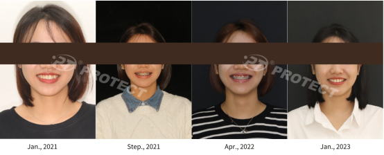
Front View Photo Comparison During Treatment
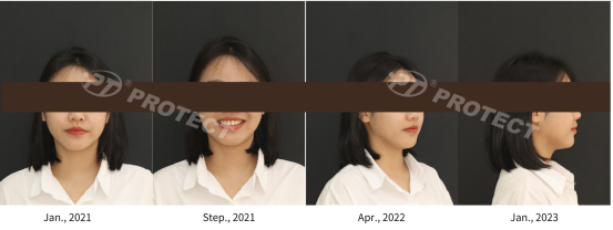
Side View Photo Comparison During Treatment
Intraoral Before-and-after Comparison
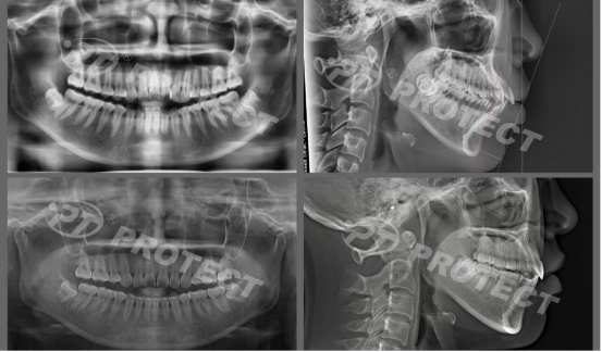
X-ray Comparison Before and After
Summary and Experience Sharing
Treatment Experience: This case primarily involved a skeletal Class II malocclusion with maxillary protrusion, mandibular retrusion, and maxillary anterior teeth overjet of more than 2mm beyond the lower lip. The use of implant anchorage screws assisted in intruding the maxillary anterior teeth. The treatment focused on controlling the occlusal plane, resulting in a 1.1° reduction in the ANB angle, achieving a counterclockwise rotation of the mandible, reducing excessive tension in the chin muscles, and improving the soft tissue profile of the chin area.
At the end of the treatment: Molar and canine teeth exhibit a Class I relationship, achieving a tight and well-aligned bite with excellent contact between adjacent teeth. The overbite measures 2mm, while the overjet is 1mm. The midlines are perfectly aligned.
The panoramic X-ray reveals nearly parallel root alignment, good molar width, a straight facial profile, a well-formed chin, and no significant joint abnormalities.
RELATED NEWS
-

-
-
Tips On Choosing and Using Orthodontic Rubber Chainsnews | 2023-12-18
We are at your service.
Whether you have inquiries about our products or need assistance with troubleshooting, our team of sales and service experts is here to assist you.
GET A QUOTE